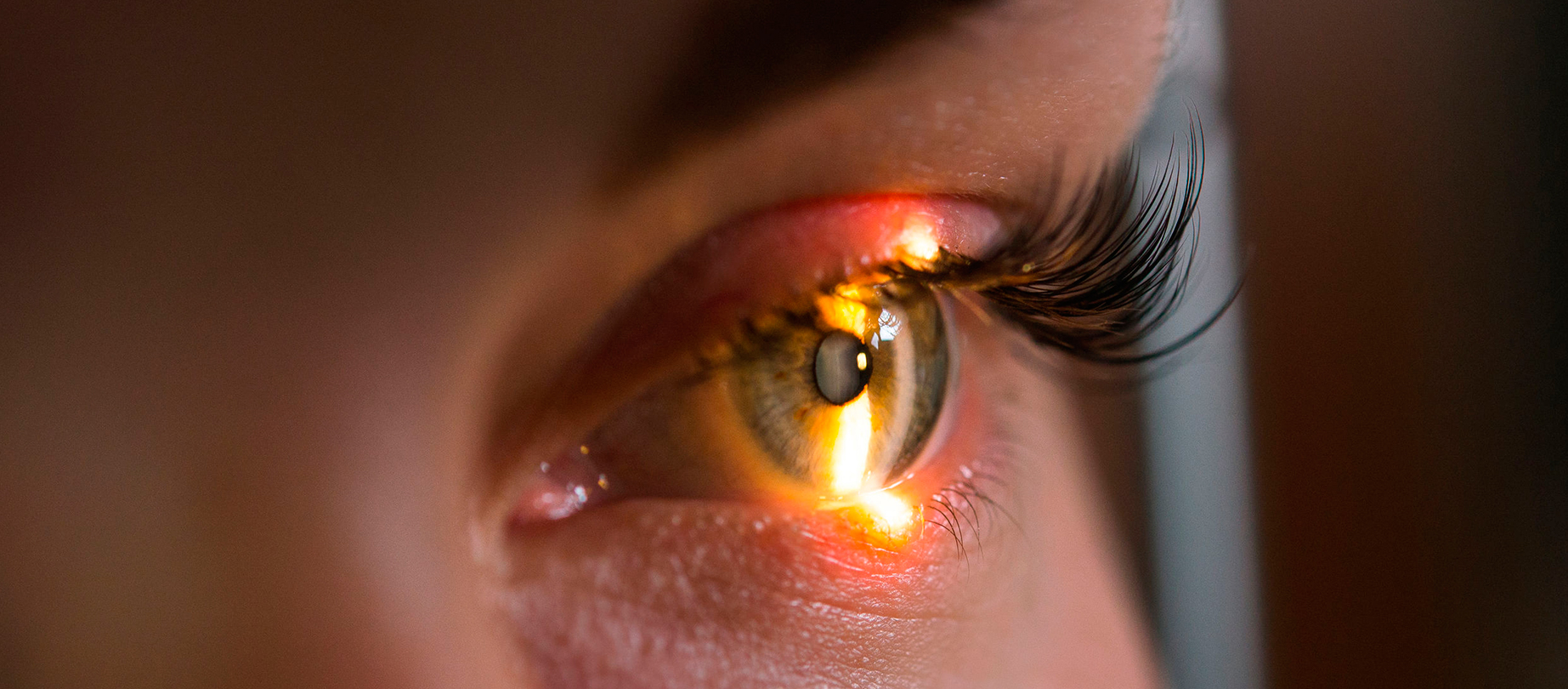
What is Keratoconus?
Keratoconus is a degenerative disease that manifests itself by progressive deformation and thinning of the cornea. It usually appears in adolescence, affects both men and women, and progresses into adulthood. Symptoms include decreased visual acuity, blurred vision, the appearance of cylinder diopters, hyperlacrimation, red eye, pain, and can progress to complete vision loss, when corneal transplant is required. The sooner it is diagnosed, the sooner it can be treated.
- Keratoconus is characterized by thinning of the cornea and the appearance of irregularities on its surface. The middle layer is the thickest part of the cornea, made mostly of water and a protein called collagen. Collagen makes the cornea strong and flexible and helps maintain its regular, round shape. In cases of keratoconus, the cornea thins and swells into an irregular cone shape, resulting in decreased vision.
- Keratoconus generally begins at puberty and progresses in the 30-40 age range. There is no way to predict how quickly the disease will progress or whether it will progress. Keratoconus usually affects both eyes, with one more severely affected than the other.
About cornea
The cornea is the surface layer of the eyeball. It is perfectly transparent and has a dome shape specific to the eye, covering its anterior surface. It comes into direct contact with the tear film and the external environment when the palpebral fissure is open.
The main role of the cornea is to refract light and obtain a clear image. Together with the lens, they are responsible for focusing light on the retina. When light hits the surface of the cornea, it refracts to pass further through the lens. The process is similar to that of a camera (the cornea and lens work as a lens system, while the retina is the image carrier, in the case of the camera, being the film or digital sensor). To maintain this role, the cornea must remain transparent and stable in shape. Any disorder that alters transparency or shape will prevent light rays from passing through to form the image. It is also a barrier against micro-organisms, dust particles or other factors that can damage the eyeball. It is avascular (no blood vessels) and has a laminar structure with layers arranged from the outside inwards.
What symptoms do you experience in case of Keratoconus?
Many keratoconus patients do not know they have the disease. The first symptom is a slight blurring of vision or progressively poor vision, which is not easily corrected. Other symptoms of keratoconus include:
- Glow and halos around lights
- Difficulty seeing at night
- Eye irritation or headaches associated with eye pain
- Increased sensitivity to bright light
- Sudden worsening or distortion of vision
What are the risk factors for Keratoconus?
The definitive cause of keratoconus is unknown, although the predisposition to develop the disease is thought to be present at birth. A common finding in keratoconus is loss of collagen in the cornea. This may be caused by some imbalance between the production and destruction of corneal tissue by corneal cells.
The following causes may increase the risk of developing keratoconus:
- Genetics. Patients with a family history of keratoconus or with certain systemic disorders, such as Down’s syndrome, have a higher risk of developing keratoconus.
- Chronic eye inflammation. Constant inflammation due to allergies or irritants can contribute to the destruction of corneal tissue which can lead to the development of keratoconus.
- Eye rubbing. Chronic eye rubbing is associated with the development of keratoconus. It may also be a risk factor for disease progression.
- Age. Keratoconus is often discovered in adolescence. In general, young patients with advanced keratoconus are more likely to need some form of surgery as the disease progresses.
What types of Keratoconus are there?
There are four main types of keratoconus, in addition to the rarer variations of the disease, including round cone, oval cone, frustule forms and keratoglobus. The shape of the cornea as well as where the cornea has become thinner determines the type of keratoconus. The type of keratoconus must be diagnosed with the help of specialist investigations and ophthalmological examination.
What treatment methods exist for Keratoconus?
In addition to a complete medical history and ophthalmologic examination, your ophthalmologist may perform the following tests to diagnose keratoconus:
- Corneal topography. This is the most accurate way to diagnose early keratoconus and track its progression. With the help of the topographer, a computerized image is made which creates a map of the corneal curve.
- Slit lamp examination. This examination of the cornea can help detect abnormalities in the outer and middle layers of the cornea.
- Pachymetry. This test is used to measure the thinnest areas of the cornea.
- Specular microscopy
- Keratometry
The treatment of keratoconus focuses on correcting vision and depends on the stage of the disease.
After investigations, the specialist doctor will determine how far the disease has progressed, whether it is progressive or stable.
Patients under the age of 40 require treatment as soon as possible.
Patients over the age of 40 require treatment, but the disease is already naturally stabilized with advancing age.
First stages
Treatment for keratoconus in the early stages includes glasses to treat myopia and astigmatism. As keratoconus progresses and worsens, glasses are no longer able to provide clear vision and patients must wear contact lenses, usually hard contact lenses.
Intermediate stages
Progressive keratoconus can be treated with surgery Crosslinking for keratoconus. The method used is photooxidative crosslinking with riboflavin by the transepithelial process, which can be performed in our clinics. This unique procedure involves applying a vitamin B2 solution to the eye, which is then activated by ultraviolet light. The solution causes new collagen bonds to form, recovering and preserving some of the strength and shape of the cornea.
Although treatment cannot make the cornea completely normal again, it can prevent vision from getting worse and, in some cases, improve vision. The procedure may require removal of the thin outer layer of the cornea (the epithelium) to allow riboflavin to penetrate the corneal tissue more easily.
Advanced stages
Corneal ring. In severe keratoconus, standard contact lenses may become too uncomfortable to wear. Intacs are implantable, plastic, C-shaped rings that are used to flatten the surface of the cornea, allowing an improved vision. They can also allow a better fit for contact lenses. The procedure takes about 15 minutes.
Corneal transplant. In a corneal transplant, a donor cornea replaces the patient’s damaged cornea.
References:

Nystagmus is the eye condition where the eyes make repetitive and uncontrollable movements. Discover other symptoms and treatment options.

Color vision deficiency, also known as dyschromatopsia, is a general term referring to various vision disorders characterized by a deficiency in color perception.

“Flying flies” are most often harmless and represent a normal stage in the aging process. Find out what the causes are and how you can reduce the symptoms.

Ocular allergies occur as a reaction of the body to an allergen, causing inflammation and itching in the eyes. The most common ocular allergies are seasonal.

Ophthalmic migraine is most common in the 40s. It manifests itself in visual impairment and even temporary blindness.

Keratitis, also known as “corneal ulcer”, is an inflammation of the cornea. If detected early, the ophthalmological disorder is easy to treat and heals quickly.
Diplopia is an ophthalmological disease in which you see two images of the same thing. The condition can affect anyone, but is more common after the age of 60.

Xanthelasma is a member of the xanthomas family and represents fatty deposits in the skin cells around the eyes. It is visible as yellow, harmless bumps.

Colorblind people perceive colors differently from most people. Most of the time, this ophthalmological disorder makes it difficult to distinguish between certain colors.

Epiphora is an ophthalmological disorder manifested by excessive tearing of the eyes. Most of the time, it is not severe and disappears on its own. However, if you are experiencing this and the problem persists, we recommend that you make an appointment for an ophthalmological examination. Treatment can be different, depending on the cause of the epiphora.

If you notice a yellow spot on the white of your eye, you are most likely dealing with pinguecula. The ophthalmological disorder is not severe, but the symptoms can be uncomfortable. Find out how to treat pinguecula and, more importantly, how you can prevent it.

Entropion is the ophthalmological disorder in which the eyelid of the eye turns inwards. It is different from ectropion, where the eyelid turns outwards. It most often occurs in older people and usually only affects the lower eyelid.

It is possible that you may also be experiencing ocular toxoplasmosis without knowing it. This retinal disorder is extremely common in our century and is manifested by eye discomfort and blurred vision.

Ectropion is the ophthalmic disorder in which the eyelid and eyelashes pull away from the cornea, and reorient outwards.

One of the most common types of headache is headache of ocular origin. It occurs as a result of an ophthalmological disorder.

Blepharitis is an ophthalmological disorder that manifests itself by inflammation of the eyelids. At the base of the eyelids, the patient notices small crusts formed by solidified oil particles or bacteria that collect in the crease at the corner of the eye.

Uveitis is an ophthalmological disorder of the uveal tract, manifested by changes in vision and eye pain.

Among the most common ophthalmological disorders is hordeolum. This is popularly known as an “stye” and is an infection of the eyelids.

The drooping eyelid is known in medical terms as “palpebral ptosis”. It manifests itself by narrowing the visual slit of one or both eyes, creating aesthetic and functional discomfort.

Amblyopia is a vision problem, popularly known as “lazy eye”. This disorder can occur in one or both eyes, and studies show that around 3% of the population suffer from this eye disease.

The sensation of “dry eyes” or “tired eyes” is known in medical terms as “dry keratoconjunctivitis” or “xerophthalmia”, and refers to a dysfunction of the tear film.

Strabismus, also known as “crossed-eyes” or “crossed vision”, is an ophthalmological disorder in which the visual axis of the eyes is not aligned. This causes one eye to deviate when it needs to look at a fixed point.

Conjunctivitis is one of the most common ophthalmological disorders. It can occur in adults, children and babies.

Chalazion is manifested by inflammation of the upper or lower eyelid. It is one of the most common ophthalmological disorders, and occurs when the secretion of sebaceous glands in the eye is blocked.

Macular degeneration involves deterioration of the macula and therefore of the quality of central vision. This disease does not affect peripheral vision and therefore cannot lead to complete blindness.

Hypermetropia affects the ability to see nearby objects. You may be able to see distant objects clearly, but closer objects, even words in a book, are usually out of focus. Hypermetropia occurs when the eye does not focus light properly on the retina (the light-sensitive layer at the back of the eye).

Myopia is a disorder that falls into the category of refractive errors (along with astigmatism and hypermetropia). In common terms, myopia manifests itself as blurred distance vision, while near vision is not a problem.

Astigmatism, like myopia and hypermetropia, is a refractive error. In general terms, the disorder manifests itself in blurred, fuzzy vision, regardless of the distance to objects, surfaces.

Presbyopia is an age-related disorder characterized by decreased near vision. It usually appears around the age of 40.

Cataract is a common ophthalmological disorder that causes progressive loss of vision through loss of lens transparency. Studies show that about 50% of the population loses their vision due to this disorder.