What is hypermetropia?
Hypermetropia affects the ability to see nearby objects. You may be able to see distant objects clearly, but closer objects, even words in a book, are usually out of focus. Hypermetropia occurs when the eye does not focus light properly on the retina (the light-sensitive layer at the back of the eye). Severe hypermetropia can make everything appear blurry, regardless of distance.
Who is at risk of hypermetropia?
Hypermetropia often affects adults over 40, but it can affect people of all ages – including babies and children. In children hypermetropia is usually genetic. Babies and young children can suffer from hypermetropia, but this should eventually correct itself as children get older. However, it can happen that one eye becomes lazy (amblyopia). This is because the eye with the poorest vision is ignored by the brain and does not learn the correct way to see. If this is not corrected in young children, there is a risk that the weaker eye will never see as well as the other eye.
What are the main symptoms of hypermetropia?
Symptoms vary depending on the age of the patient and the severity of the refractive error. The patient may be asymptomatic. Low refractive error in young patients is usually corrected by mild accommodative effort without producing symptoms. But if the eye muscles have to work harder, you may develop symptoms such as:
- Some people have trouble just focusing on nearby objects, while others can struggle to see clearly at any distance.
- You find that nearby objects appear blurred and out of focus, but distant objects are clear
- You have to squint to see clearly
- Have tired or strained eyes after activities that involve focusing on nearby objects, such as reading, writing or working on a computer
- Headaches
- Slight aversion to light/photophobia
Children with hypermetropia often don’t initially have obvious problems with their vision. But if left untreated, it can lead to problems such as the lazy eye.
What causes hypermetropia?
Eyes affected by hypermetropia do not refract (bend) light properly. They under-focus light and form an incomplete image on the retina. When this happens, you cannot see clearly.
To make it easier to understand, consider how normal vision works.
- The light penetrates the eyes.
- Light refracts when it passes through two parts. First the cornea, the covering at the front of the eye, then the lens, a clear part that focuses light deeper into the eye.
- Light forms a focused spot on the retina, a thin layer of tissue at the back of the eye
- The retina sends information to the brain.
- The brain translates messages into images.
Hypermetropia occurs when there is a problem with the second step, the refraction.
This may be because:
- The eyeball is too short. This means that the retina is closer to the pupil, causing light to pass past the retina. A normal eye is usually about 23 mm long, so a hypermetropic eye will be shorter than 23 mm.
- The cornea (the transparent layer at the front of the eye) is too flat. The cornea needs to be curved to direct light to the retina.
- The lens inside the eye cannot focus properly
Sometimes hypermetropia can be the result of genes you inherited from your parents or a lens in your eyes that is more rigid and less able to focus as you age.
How can Hypermetropia be prevented?
There’s no proven way to prevent hypermetropia, but you can keep your eyes healthier by taking a few steps:
- A proper diet: dark green leafy greens are good for your eyes, as are fish rich in omega-3 fatty acids.
- Get regular ophthalmological examinations: an ophthalmologist can check for eye problems before you have symptoms.
- Wear sunglasses, even on cloudy days – choose sunglasses that block 99% or more of the sun’s ultraviolet (UV) radiation.
- Rest your eyes regularly: looking at a computer or reading for long periods of time can tire your eyes. Every 20 minutes, look at something far away for 20 seconds.
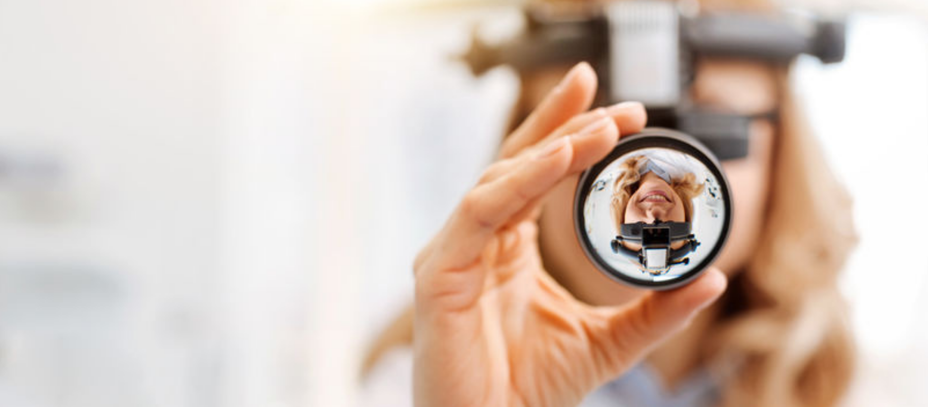
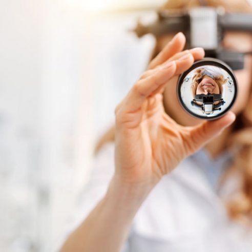
What investigations are needed for hypermetropia?
Hypermetropia is detected during an ophthalmological examination. It is usually recommended to have an examination at least once every 2 years, but you can have a test at any time if you have concerns about your vision.
Ophthalmological examinations are painless and may involve:
- Eye drops: Special eye drops can be used to dilate your eyes. The drops will increase the size of the pupils (the black center of the eye) to let in more light and allow the ophthalmologist to examine the retina.
- Autorefractometry: This instrument measures the refractive error or severity of the problem. The instrument looks like a large frame with lenses. It helps your ophthalmologist determine how to correct your vision.
- Biomicroscopy: Your ophthalmologist will focus a special light into your eyes to see how it reflects in the retina. This step helps you determine if you are farsighted or nearsighted. This isn’t commonly done, except in pediatric cases (for children).
How is Hypermetropia treated?
Children and young adults with hypermetropia may not need any treatment, as their eyes are often able to adapt to the problem and their vision may not be significantly affected.
Treatment is usually necessary in older adults, especially those over 40, because the eyes become less able to adjust as they age.
There are several ways in which hypermetropia can be corrected. The main treatments are:
- prescription glasses – these have lenses designed specifically for you, which ensure that the light is focused correctly on the back of your eyes
- contact lenses – some people prefer them to glasses because they are lightweight and virtually invisible
- laser surgery – the laser is used to change the shape of the cornea, which can mean you don’t have to wear glasses or contact lenses
- Lens implant surgery
Glasses are the simplest and safest treatment. Contact lenses and laser surgery have a low risk of complications and are not usually indicated for children.
Hypermetropia does not go away unless you have surgery. Even with surgery, you may find that the disorder returns after a few years.
With glasses and contact lenses, your vision can improve and fade over time. It is important to wear glasses or contact lenses whenever your eye doctor recommends it. You should also have regular ophthalmological examinations if you need to change your lens diopters.
What surgeries can be performed for hypermetropia?

Nystagmus is the eye condition where the eyes make repetitive and uncontrollable movements. Discover other symptoms and treatment options.

Color vision deficiency, also known as dyschromatopsia, is a general term referring to various vision disorders characterized by a deficiency in color perception.

“Flying flies” are most often harmless and represent a normal stage in the aging process. Find out what the causes are and how you can reduce the symptoms.

Ocular allergies occur as a reaction of the body to an allergen, causing inflammation and itching in the eyes. The most common ocular allergies are seasonal.

Ophthalmic migraine is most common in the 40s. It manifests itself in visual impairment and even temporary blindness.

Keratitis, also known as “corneal ulcer”, is an inflammation of the cornea. If detected early, the ophthalmological disorder is easy to treat and heals quickly.
Diplopia is an ophthalmological disease in which you see two images of the same thing. The condition can affect anyone, but is more common after the age of 60.

Xanthelasma is a member of the xanthomas family and represents fatty deposits in the skin cells around the eyes. It is visible as yellow, harmless bumps.

Colorblind people perceive colors differently from most people. Most of the time, this ophthalmological disorder makes it difficult to distinguish between certain colors.

Epiphora is an ophthalmological disorder manifested by excessive tearing of the eyes. Most of the time, it is not severe and disappears on its own. However, if you are experiencing this and the problem persists, we recommend that you make an appointment for an ophthalmological examination. Treatment can be different, depending on the cause of the epiphora.

If you notice a yellow spot on the white of your eye, you are most likely dealing with pinguecula. The ophthalmological disorder is not severe, but the symptoms can be uncomfortable. Find out how to treat pinguecula and, more importantly, how you can prevent it.

Entropion is the ophthalmological disorder in which the eyelid of the eye turns inwards. It is different from ectropion, where the eyelid turns outwards. It most often occurs in older people and usually only affects the lower eyelid.

It is possible that you may also be experiencing ocular toxoplasmosis without knowing it. This retinal disorder is extremely common in our century and is manifested by eye discomfort and blurred vision.

Ectropion is the ophthalmic disorder in which the eyelid and eyelashes pull away from the cornea, and reorient outwards.

One of the most common types of headache is headache of ocular origin. It occurs as a result of an ophthalmological disorder.

Blepharitis is an ophthalmological disorder that manifests itself by inflammation of the eyelids. At the base of the eyelids, the patient notices small crusts formed by solidified oil particles or bacteria that collect in the crease at the corner of the eye.

Uveitis is an ophthalmological disorder of the uveal tract, manifested by changes in vision and eye pain.

Among the most common ophthalmological disorders is hordeolum. This is popularly known as an “stye” and is an infection of the eyelids.

The drooping eyelid is known in medical terms as “palpebral ptosis”. It manifests itself by narrowing the visual slit of one or both eyes, creating aesthetic and functional discomfort.

Amblyopia is a vision problem, popularly known as “lazy eye”. This disorder can occur in one or both eyes, and studies show that around 3% of the population suffer from this eye disease.

The sensation of “dry eyes” or “tired eyes” is known in medical terms as “dry keratoconjunctivitis” or “xerophthalmia”, and refers to a dysfunction of the tear film.

Strabismus, also known as “crossed-eyes” or “crossed vision”, is an ophthalmological disorder in which the visual axis of the eyes is not aligned. This causes one eye to deviate when it needs to look at a fixed point.

Conjunctivitis is one of the most common ophthalmological disorders. It can occur in adults, children and babies.

Chalazion is manifested by inflammation of the upper or lower eyelid. It is one of the most common ophthalmological disorders, and occurs when the secretion of sebaceous glands in the eye is blocked.

Macular degeneration involves deterioration of the macula and therefore of the quality of central vision. This disease does not affect peripheral vision and therefore cannot lead to complete blindness.
Hypermetropia affects the ability to see nearby objects. You may be able to see distant objects clearly, but closer objects, even words in a book, are usually out of focus. Hypermetropia occurs when the eye does not focus light properly on the retina (the light-sensitive layer at the back of the eye).

Myopia is a disorder that falls into the category of refractive errors (along with astigmatism and hypermetropia). In common terms, myopia manifests itself as blurred distance vision, while near vision is not a problem.

Astigmatism, like myopia and hypermetropia, is a refractive error. In general terms, the disorder manifests itself in blurred, fuzzy vision, regardless of the distance to objects, surfaces.

Presbyopia is an age-related disorder characterized by decreased near vision. It usually appears around the age of 40.

Cataract is a common ophthalmological disorder that causes progressive loss of vision through loss of lens transparency. Studies show that about 50% of the population loses their vision due to this disorder.